All three kinds of exercise resulted in an increase of beta activity in Brodmann area 7. 

Animal experiments showed that the parietal cortex is also important for "counting behavior" in monkeys (area of the intraparietal sulcus and area 5), dogs (area 5) and cats (area 7).
The subjective pleasantness and richness were modulated by the word labels, as were the fMRI activations to touch in parietal cortex area 7, the insula and ventral striatum.
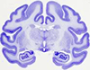
Anatomically, the precuneus corresponds to the medial portion of human cerebral cortical Brodmann area 7.
RESULTS: Age of onset showed a positive correlation with regional gray matter volume in the right superior parietal lobule (Brodmann area 7).
We establish that ordered patterns activated only primary visual cortex - V1 and V2, (BA17-18), wheareas chaotic patterns activated in addition primary visual cortex, the V3,V4,V5 (BA19) of the occipital cortex and the area 7 of parietal area (BA7) classification.
Wheat germ agglutinin horseradish peroxidase (WGA-HRP) was administrated by micro-electrophoresis and micro-injection, respectively, into area 17 and area 7 in different hemispheres in eight cats. After such restricted injections labeled pyramidal cells were observed in layer 5 of area 7. These pyramidal cells were arranged as discontinuous patches extending across a broad region of area 7. These results suggest that feedback from area 7 to area 17 may arise from specific functional columns in area 7..
In non-human primates (monkeys), it has been reported for the primary sensory cortices (A1, V1, S1), the motor and premotor cortical areas and, in the parietal lobe, also for area 7.
Combined bilateral lesions of areas 5 and 7 and an isolated lesion of area 5 resulted in a severe impairment of the numerical discrimination for two months, whereas the isolated lesion of area 7 did not lead to any problems in differentiation.
Forebrain ischemia-reperfusion neural injury in rats was demonstrated by histopathological observation, which revealed significant neural cell death in the hippocampus CA1 area 7 days post-ischemia (77% cell loss).
In a previous study, we identified three cortical areas in human posterior parietal cortex that exhibited topographic responses during memory-guided saccades [ visual area 7 (V7), intraparietal sulcus 1 (IPS1), and IPS2], which are candidate homologs of macaque parietal areas such as the lateral intraparietal area and parietal reach region.
The inferior parietal lobule (IPL) of the macaque monkey constitutes the largest part of Brodmann's area 7. Functional, connectional, and architectonic data have indicated that area 7 is comprised of several distinct sectors located in the lateral bank of the intraparietal sulcus and on the IPL cortical convexity.
We describe the organization of the dorsolateral frontal areas in marmoset monkeys using a combination of architectural methods (Nissl, cytochrome oxidase, and myelin stains) and injections of fluorescent tracers in extrastriate areas (the second visual area [ V2], the dorsomedial and dorsoanterior areas [ DM, DA], the middle temporal area and middle temporal crescent [ MT, MTc], and the posterior parietal cortex [ area 7]). The intermediate subdivision of area 8 (8Ad) has efferent projections to area 7, while the dorsomedial subdivision (8B) has few or no connections with extrastriate cortex. Area 46, located rostrolateral to area 8Av, has substantial connections with the medial extrastriate areas (DM, DA, and area 7) and with MT, while the cortex lateral to 8Av (area 12/45) projects primarily to MT and to the MTc. Finally, cells in dorsal area 6 (6d) have sparse projections to DM, MT, and the MTc, as well as strong projections to DA and to area 7.
Only parietal area 7 bilaterally was non-negligibly active in all subjects (currents above 10% maximum). Based on the present results, the concept of a universal attention-related set of cortical areas if restricted to common areas across subjects is challenged, since even area 7 may no longer be common when the sample size becomes larger.
We concentrate on six of these: the frontal eye field, parietal eye field, supplementary eye field, middle superior temporal area, prefrontal eye field, and area 7 m (precuneus in humans).
Sources of these potentials were located in occipital area 19 and parietal area 7.
Administration of S34176 (75 mg/kg i.p.) 30 min before transient (10 min) global ischaemia in Wistar rats significantly prevented delayed neuronal cell death in the hippocampal CA1 area 7 days post-ischaemia (24% vs.
Corticopulvinar cells were located in layers V and VI of a wide variety of cortical areas, with a major concentration of cells in area 7. We examined corticopulvinar terminals labeled from area 7 at the ultrastructural level in tissue stained for gamma-aminobutyric acid (GABA). Interpretation of these results using Sherman and Guillery's recent theories of thalamic organization (Sherman and Guillery [ 1998] Proc Natl Acad Sci U S A 95:7121-7126) suggests that area 7 may both drive and modulate PUL activity..
We studied a patient who experienced 'palinaesthesia', an illusion of persistent touch following tactile stimulation on the left hand, subsequent to a right parietal meningioma affecting primary somatosensory regions in the postcentral gyrus (SI) and superior parietal gyrus (Brodmann area 7), but preserving the secondary somatosensory cortex (SII) in the upper lateral sulcus.
In multisensory cortex, unilateral deactivation of neither ventral or dorsal posterior ectosylvian cortices nor anterior or posterior area 7 resulted in any deficits.
Cooling deactivation of: 1) dorsal or 2) ventral posterior suprasylvian gyrus; 3) ventral posterior ectosylvian gyrus, 4) middle ectosylvian gyrus; 5) anterior or 6) posterior middle suprasylvian gyrus (area 7); 7) anterior middle suprasylvian sulcus; 8) medial area 5; 9) the visual portion of the anterior ectosylvian sulcus (AES); 10) or lateral area 6 were all without impact on the ability to redirect gaze.
By maintaining strict control over experimental conditions including duration of insult, temperature and humidity, we produced a reliable model of minor injury primarily affecting all five areas of the cerebral cortex, and also the thalamus (area 7) and basal ganglia (area 8).
A large number of imaging studies have identified a role for the posterior parietal lobe, in particular Brodmann's area 7 and the intraparietal sulcus (IPS), in mental rotation. Repetitive transcranial magnetic stimulation (rTMS) was applied to posterior parietal locations estimated to overlie Brodmann's area 7 in the right and the left hemisphere, or to a posterior midline location (sham condition).
Medium projections could be traced to the ventrolateral prefrontal and lateral orbital cortex (areas 47L and O), the primary somatosensory areas 3b and 2, the agranular and dysgranular insula, and the posteroinferior parietal cortex (area 7; PFG, PG).
Posterior cortical areas also contributed importantly to the SP, for instance extrastriate area 19 and parietal area 7, in 90% of the subjects.
Two cats sustained large unilateral ablations of the contiguous visual areas, and cooling loops were placed in the pMS sulcus, and in contact with adjacent area 7 or posterior ectosylvian (PE) cortex of the opposite hemisphere. In both instances cooling of pMS cortex, but neither area 7 nor PE, restored a virtually normal level of orienting performance to stimuli presented anywhere in the previously hemianopic field.
There is considerable evidence from studies on cats and monkeys that several cortical areas such as area 2v at the tip of the intraparietal sulcus, area 3av in the sulcus centralis, the parietoinsular vestibular cortex adjacent to the posterior insula (PIVC) and area 7 in the inferior parietal lobule are involved in the processing of vestibular information.
However, in other areas (FEF and parietal area 7), strong activation was observed even at the lowest attentional load (compared to a passive baseline using identical stimuli), but little or no additional activation was seen with increasing load.
The four regions of parietal cortex we examined included the: middle suprasylvian (MS) gyrus (area 7), anterior middle suprasylvian (aMS) sulcus (AMLS, ALLS), posterior middle suprasylvian (pMS) sulcus (PMLS, PLLS), and the dorsal posterior suprasylvian (dPS) gyrus (area 21a).
These included Brodmann areas 19/37, the inferior (Brodmann Area 39), and superior parietal lobule (Brodmann area 7).
Different subregions of the intraparietal cortical area 7 were activated by fixation, saccades to visual targets, and acoustically triggered reaching in the dark.
Pyramidal neurones were injected with Lucifer Yellow in slices cut tangential to the surface of area 7 m and the superior temporal polysensory area (STP) of the macaque monkey. Sholl analyses revealed that layer III pyramidal neurones in area STP had considerably higher peak complexity (maximum number of dendritic intersections per Sholl circle) than those in layer V, whereas peak complexities were similar for supra- and infragranular pyramidal neurones in area 7 m. Calculations of the total number of dendritic spines in the "average" basal dendritic arbor revealed that layer V pyramidal neurones in area 7 m had twice as many spines as cells in layer III (4535 and 2294, respectively).
Low-resolution electromagnetic tomography demonstrated, independent of the scalp distribution, a distributed spindle source in the prefrontal cortex (Brodmann areas 9 and 10), oscillating with a frequency below 13Hz, and in the precuneus (Brodmann area 7), oscillating with a frequency above 13Hz. Brodmann areas 9 and 10 have principal connections to the dorsomedial thalamic nucleus; Brodmann area 7 is connected to the lateroposterior, laterodorsal and rostral intralaminar centrolateral thalamic nuclei.
Because the SMG is part of area 7, which belongs to a network of multisensory visual-vestibular cortical areas, we conclude that a small lesion there can cause motion sickness susceptibility..
Subsequently, three pairs of cooling loops were implanted bilaterally in contact with visuoparietal cortices forming the crown of the middle suprasylvian gyrus (MSg; architectonic area 7) and the banks of the posterior-middle suprasylvian sulcus (pMS sulcal cortex) and in contact with the ventral-posterior suprasylvian (vPS) region of visuotemporal cortex. Bilateral deactivation of pMS sulcal cortex resulted in a profound impairment for all six tested positions of the landmark, yet bilateral deactivation of neither area 7 nor vPS cortex yielded any deficits. Therefore, we conclude that bilateral cooling of pMS cortex, but neither area 7 nor vPS cortex, induces a specific deficit in spatial localization as examined with the landmark task.
Ischemic damages, evaluated by histopathological grading of hippocampal CA1 area 7 days after ischemia, was significantly ameliorated in the mild (1.3+/-0.5, mean+/-S.E.M.) and moderate hypothermic rats (0.8+/-0.3) compared with the normothermic ones (3.4+/-0.4).
The ALS group showed statistically significant decreases in relative FMZVD in the prefrontal cortex (areas 9 and 10 bilaterally), parietal cortex (area 7 bilaterally), visual association cortex (area 18 bilaterally) and left motor/premotor cortex (including area 4) (P < 0.001).
area 7) cortical regions.
Here we investigated the relative contributions of top-down and bottom-up directed interactions between area 17 and area 7 of the cat visual system.
Stimulation of the impaired hand resulted in activation of the ipsilateral parietal operculum (second somatosensory area [ SII]) and posterior parietal lobe (Brodmann's area 7) in all cases, but no activation was elicited in the SI in any patient.
Significant activation was found in only one area located in the right posterior parietal lobe, centred on the intraparietal sulcus (Brodmann area 7).
Pyramidal neurones were injected with Lucifer Yellow in cortical slices taken from layer III of the medial subdivision of cytoarchitectonic area 7 (7m) of the macaque monkey. Layer III pyramidal neurones in area 7m have an average basal dendritic field area of 109.57 +/- 13.03 x 10(3) microm2, which is significantly greater than that obtained for neurones in the lateral intraparietal area (LIP) and area 7a. Moreover, Sholl analyses revealed that neurones in area 7m are significantly more complex in their branching patterns than those in LIP and area 7a. These results reinforce the view that, behind the apparent architectural uniformity of Brodmann's area 7, there is a significant diversity of neuronal structure and function..
fMRI demonstrated bilateral activation in the superior parietal lobulus (Brodmann area 7) with a right/left ratio of 1.95.
Extirpation of the parietal cortex area 7 aggravated delayed visual discrimination of all visual attributes including shape, colour and spatial relationship in adult rhesus monkeys.
The ipsilateral premotor area (Brodmann area 6), bilateral posterior parietal areas (Brodmann area 7) and precuneus showed an increase in rCBF related only to the length of the sequences, without any change from rest to simple repetitive movement.
Positive correlations of rCBF with increasing sequence complexity were identified in the contralateral rostral supplementary motor area (pre-SMA) and the associated pallido-thalamic loop, as well as in right parietal area 7 and ipsilateral primary motor cortex (M1).
Two cortical areas in rats have been found to be important in directed attention and spatial processing: the medial agranular cortex (AGm), the rodent analog of the frontal eye fields; and the posterior parietal cortex (PPC), the rodent analog of area 7 in primates.
The role of area 7 m has been studied by recording the activity of single neurons of monkeys trained to fixate and reach toward peripheral targets. The results show that cell activity in area 7 m relates, for some cells to eye position, for others to hand position and movement, and for the majority of cells to a combination of visuomanual and oculomotor information.
Many neurons in the human somatosensory cortex (area 7 of Brodmann) possess an intensely negative-charged surface coat consisting of perineuronal sulfated proteoglycans which were stained with cationic iron colloid.
The right dorsal premotor cortex (Brodmann area 6) and the right precuneus (Brodmann area 7) showed a linear increase of rCBF as sequence complexity increased.
Lesions of the posterior parietal system in humans and of the presumably homologous area 7 of the monkey cause impairment in high-order manipulative behavior. To clarify the nature of normal functioning in this domain, a study was done by microelectrode recording from area 7 of the monkey of neural firing in relation to hand reaching to targets. The data obtained show that an area 7 neuron fires in relation to simple stimuli, such as a square of white light, and to composite stimuli, such as hand reaching by the monkey or by the trainer in the monkey's extrapersonal space, the response to complex stimuli characteristically multiduty. It is proposed that functional linkage of cognate features, a consequent modular category encoding and "packaging" of information relevant for praxis schema, is thus achieved by area 7 cells specialized for integrating information in the time and space domains visual, tactile, proprioceptive/kinaesthetic, and other for the sake of behavior in extrapersonal space.
On the other hand, the posterior parietal association cortex (Brodmann's area 7) was more activated in the finger, direction, and none conditions than in the full condition.
The anterograde tracer 3H-leucine was pressure injected bilaterally into the cortex of six monkeys (for a total of 12 cases) involving the primary visual cortex (area 17); the medial prestriate cortex (medial 18/19); dorsomedial area 19; the caudal portion of the cortex of the superior temporal sulcus, upper bank (cytoarchitectural area OAa) and lower bank (area PGa); the lower bank of the caudal lateral intraparietal sulcus (area POa); and the inferior parietal lobule (area 7). The posterior pretectal nucleus received sparse projections from area 7 and the cortex lining the intraparietal sulcus (dorsomedial part of area 19 and POa). The pretectal olivary nucleus was targeted by neurons in cortex of dorsomedial area 19, and the anterior pretectal nucleus was targeted by neurons in both dorsomedial 19 and area 7. Rostral area 7 (mainly 7b) neurons terminated in the stratum album intermediale (SAI); no SC terminals were found in a case in which caudal area 7 (mainly 7a) was injected..
Compared to the relaxed state, task performance was distinguished by a drop in power for frequencies below 20 Hz (most prominent in area 7), and an increase for frequencies above 20 Hz.
Also, four zones that were free of callosal connectivity in area 7, on the banks of the suprasylvian sulcus, and in the posterior suprasylvian sulcus were found in both normal and enucleated cats.
The lesion involved area 5, parts of area 7, the angular gyrus, the middle and posterior parieto-occipital gyri, and posterior parts of the superior and middle temporal gyri.
Both the coherent and incoherent movements activated a part of the superior parietal lobule located within the intraparietal sulcus (Brodmann area 7).
Less abundant but consistent projections were detected in cingular, auditory II, lateral suprasylvian and anterior ectosylvian visual cortices, and cortical area 7.
These were located (i) bilaterally in the precuneus of superior parietal cortex (area 7 of Brodmann); (ii) bilaterally in the cuneus (a region considered to represent upper V3); (iii) in the left lingual and fusiform gyri (possibly lower V3 and adjacent areas).
Whereas in humans Brodmann's area 7 is above this sulcus, in monkeys it is below and therefore part of the IPL. Some investigators contend that the monkey homologue of the human IPL (areas 39 and 40) is the monkey's IPL (area 7).
On MRI a post-traumatic porencephalic lesion was seen in area 7 and the superior part of area 39 of Brodmann; on T2 sequences, it was surrounded by a hyperecho predominating in the inferior part of the parietal lobe and extending in the posteroexternal temporal cortex.
We obtained visual response latencies and response durations following visual stimulation of neurones recorded in the lateral geniculate nucleus (LGN), areas 17, 18, 19, the posteromedial lateral suprasylvian sulcus (PMLS), and area 7 of the cat.
Only a few efferent connections to the brainstem vestibular nuclei were found for the different parts of cytoarchitectonic area 7.
Transient staining in extrastriate visual cortical areas disappeared first from the lateral suprasylvian areas, and persisted longest in area 7.
We suppose that the parietal lobe, especially Broadmann's area 7 may be one of the responsible areas for provoking hemiballistic involuntary movement..
Our results give support to a functional segregation between the lateral suprasylvian visual areas (LSA) and area 7. After lesion of area 7, binocular depth cues can no longer be used.
This research is focused on the contribution of area 7 to the short-term visual spatial memory. In the second stage, bilateral area 7m was lesioned.
In the superior parietal lobule (area 7) of AIDS brains, no loss of nerve cells was noted.
The contribution of the lateral suprasylvian cortex to pattern recognition was studied by behavioural detection experiments in combination with bilateral lesions of different parts of the lateral suprasylvian areas (LSA) and area 7 in seven cats. Four different types of lesion could be distinguished depending on their extent: (1) lesion of parts of the (LSA); (2) lesion of parts of the LSA with undercutting of areas 17, 18 and 19; (3) lesion of area 7; (4) lesion of area 7 and parts of the LSA.
In control experiments thalamic projections to the granular insula Ig and the anterior part of area 7, two cerebral structures connected with the vestibular cortical areas, were studied.
Small iontophoretic or pressure injections of horseradish peroxidase (HRP), wheat-germ-HRP, Nuclear Yellow, and Fast Blue were administered to the cytoarchitectonic areas Ri (PIVC), 3aV, the parieto-temporal association area T3, the granular insula (Ig), and the rostral part of area 7 (7ant). Area T3 receives signals from the insular and retroinsular cortex, various parts of area 7, visual areas of the parieto-occipital and parieto-temporal regions (area 19) and from a sector of the upper bank of the temporal sulcus (STS-area).
There was no difference in the binding between the amyotrophic lateral sclerosis cases and the controls in area 7 of the occipital cortex, an area which is relatively spared in amyotrophic lateral sclerosis..
When hooded rats with bilateral lesions of Krieg's area 7 (parietal cortex) were trained to locomote toward visual targets in a runway, they ran less accurately than did controls, although unilaterals ran accurately.
Rats with bilateral lesions of posterior parietal cortex (PPC: Krieg's area 7) or dorsal hippocampus (HIP) were compared with controls for their response to environmental change.
Neuronal responses of area 7 of the parietal associative cortex (PAC) to paired stimulation of the nLD and nLP thalamic nuclei were investigated by extra- and intracellular recording methods.
The distribution within individual cytoarchitectonic areas of the cells of origin of ipsilateral cortico-cortical fibres to area 7 of the parietal lobe of the monkey has been studied. After injections of horseradish peroxidase into area 7, labelled cells in a variety of cortical areas were plotted and their distribution along the length of the cortex analysed.
In contrast, when WGA-HRP was injected into the PMv immediately caudal to the arcuate sulcus and lateral to the spur of the arcuate sulcus, the labeled cells were found in area 7 (areas POa, PF, PFG), area 5 (area PEa), area PFop (secondary somatosensory area), SMA, the cingulate cortex (areas 24), caudal region of area 4 in the rostral bank of the central sulcus, and area 3a.
We were able to identify regions in Galago which resemble Macaca posterior parietal area 7, superior temporal polysensory cortex (ST), inferotemporal visual cortex (IT), the temporoparietal auditory area (Tpt), and posterior parahippocampal cortex (areas TH and TF). area 7, ST, and IT can each be subdivided further in Macaca, and for most of these subdivisions we were able to identify counterparts in Galago.
Afferent connections of LP of the thalamus and parietal area 7 from the retrosplenial cortex of rat were investigated by means of the HRP retrograde transport. It was shown that neurons of area 29d sent afferent fibres to LP, while area 7 received axons from cells of areas 29d and 29c. Apart from the neocortical input area 7 received afferent fibres from the archicortex.
Influence of external stimuli and food motivation on neuronal spike responses (area 7) induced by conditional and unconditional stimulation were studied in the awake cats.
In order to assess the relative importance of visual input to area 7 reach-related neuronal activity, a monkey was trained to reach to visual targets displayed on a video-monitor, both with and without visual feedback. Of 19 reach-related cells recorded in area 7 both in the light and the dark, ten showed an enhancement of discharge in the dark. These included area 7b cells sensitive to screen contact and area 7a cells active during reach.
As the injection site was advanced into the dorsal genu, the labeled region shifted dorsally toward the parietal lobe, including prestriate areas MT and PO, parietal area PG (Brodmann's area 7), the ventral and lateral intraparietal sulcal areas (VIP and LIP, respectively), and area PE and adjacent area LC (Brodmann's areas 5 and 23, respectively).
The corticocortical connections between area 7 and the frontal lobe have been studied in the monkey. Injections of HRP were made into area 7 of the parietal lobe or into area 46 in the walls of the principal sulcus. The two subdivisions of area 7, 7a or PG and 7b or PF, are connected with different parts of the frontal lobe, and each subdivision is connected with two distinct areas. area 7b, PF, is connected in a well organized and somatotopic manner with the lower premotor area and with the lower part of area 46, below the fundus of the principal sulcus. area 7a, PG, is connected with area 8a and with the upper part of area 46, above the fundus of the principal sulcus; it is suggested that the lower part of area 8a and the posterior part of area 46 are related to the central visual field, while the medial part of area 8a and the anterior part of area 46 are related to the periphery of the visual field. The corticocortical connections between area 7 and the frontal lobe are reciprocal and those passing from area 7 to the frontal lobe are 'feed-forward' and those to area 7 are 'feed-back'..
Spike activity of neurons (area 7) was studied during conditioned placing reflex in the rest, during appearance and extinction of the orienting reaction in trained cats.
Light-flash stimulation evoked responses with the shortest latencies in area 7.
area 7 projects to the posterior part of the intermediate and posterior suprasylvian sulcus belt. area 7 is connected only with the superior extremity of the middle ectosylvian gyrus or of areas 22, 50..
Projections of the thalamic neurons to the visual (area 17) and the parietal association (area 7) cortices were examined by retrograde axonal transport of fluorescent dyes. It was found that the pulvinar neurons can be divided into three groups with respect to their connections with these cortical areas: 1--projecting to area 7 (the largest cell group); 2--projecting to area 17 (a smaller cell group) and 3--sending their axons to the both cortical areas (only few cells).
Connections of the posterior parietal cortex (area 7) with subcortical structures related to the vestibulo-ocular function were studied on four macaque monkeys by using anterograde and retrograde tracer. Wheat germ agglutinin (WGA)-horseradish peroxidase (HRP) or tritiated amino acids were injected into the posterior part of area 7, including the caudal end of the superior bank of both the superior temporal sulcus and the lateral sulcus. Through these connections, area 7 might control the vestibulo-ocular response (VOR) by modulating the ascending vestibular information. This cortical area 7 also projects to the ipsilateral intermediate and deep layers of the superior colliculus and to several ipsilateral pontine nuclei. Cortical area 7 also was seen to project to the accessory nucleus of Darkschewitsch, to all the vestibular nuclei, and to the nucleus propositus hypoglossi; the last two projections were found to be bilateral with a greater ipsilateral contribution.
Connections of parietal cortex (especially posterior part of area 7) with subcortical structures related with vestibular function were examined in four monkeys (Macaca Fascicularis), by means of anterograde labeling with HRP and tritated amino-acids tracers. These latter efferent projections of parietal cortex onto vestibular nuclei complex could account for the role played by the posterior part of area 7 in the modulation of the vestibulo-ocular reflex..
The ability of two cats to discriminate between two geometrical outline patterns in the presence of superimposed structured background was tested before and after bilateral removal of the lateral suprasylvian visual areas (PMLS, PLLS, AMLS, ALLS, part of area 7).
In recording experiments from area 7a, a cortical subdivision in the posterior parietal cortex in monkeys, we have found neurons whose responses are a function of both the retinal location of visual stimuli and the position of the eyes in the orbits. By combining these signals area 7 a neurons code the location of visual stimuli with respect to the head. To code location in craniotopic space at all eye positions (eye-position-independent coding) an additional step in neural processing is required that uses information distributed across populations of area 7a neurons. We describe here a neural network model, based on back-propagation learning, that both demonstrates how spatial location could be derived from the population response of area 7a neurons and accurately accounts for the observed response properties of these neurons..
area 7 is connected mostly with the medial part of the lower lip of the cruciate sulcus (areas 6iffu, 6aa, 6ab) and its projection to areas 4fu and 4 delta is less pronounced. No projections were found from area 7 to areas SI and SII..
Connections of the posterior parietal cortex (area 7) with the vestibular complex have been studied in 4 macaque monkeys by anterograde axonal transport methods. WGA-HRP and tritiated amino-acids have been injected in the posterior part of area 7 including the caudal end of the superior bank of superior temporal sulcus and the lateral sulcus. Two main groups of area 7 efferences were found to project to vestibular complex: a) A first group terminates on vestibular nuclei (the inferior vestibular nucleus and the caudal part of the medial nucleus) mainly connected with cerebello-spinal system. The prepositus hypoglossi nucleus has also been found to receive area 7 projections. It is concluded that the possible control played by area 7 on the vestibulo-ocular reflex might be exerted through these direct cortico-vestibular projections..
Cortical sources of input to area 6m include several retinotopically organized extrastriate visual areas (AMLS, ALLS, and PLLS), association areas with strong links to the visual system (area 7, granular insula, posterior ectosylvian gyrus, and cingulate gyrus), and a lateral division of area 6 (area 61) with oculomotor functions. Projections from area 7, the posterior cingulate area, the ventral anterior nucleus, and the mediodorsal nucleus are spatially ordered in a pattern such that parts of area 6 close to the fundus of the cruciate sulcus receive input from neurons positioned anteriorly in the cortical areas, dorsolaterally in the ventral anterior nucleus, and ventrolaterally in the mediodorsal nucleus.
The behavioral, anatomical, and electrophysiological effects of posterior parietal cortex lesions (Krieg's area 7) were compared in rats with removals at 1, 5, or 10 days of age or in adulthood.
It has been shown earlier that binocular visual deprivation during the early sensitive period of life reduces the representation of visual functions in the posterior parietal association cortex of monkeys, in Brodmann's area 7 (Exp. Moreover, the representation of somatic functions increases suggesting that competitive mechanisms between the inputs from different modalities function during the early sensitive period of life in area 7. Transdural extracellular multiunit recordings were performed in Brodmann's area 7 at the end of the deprivation period after the opening of the eyes. A second set of recordings was conducted in area 7 after a recovery period of 12 months from the deprivation. The results indicate that visual deprivation during the early critical period of life results in a profound and persistent reduction of visual functions in area 7. However, activity-dependent competition between inputs from different modalities continue, resulting in the domination of somatosensory and somatomotor functions over visual functions in area 7.
area 7 of the cat, as identified cytoarchitecturally, includes cortex both on the middle suprasylvian gyrus and on the anterior lateral gyrus. Deposits of distinguishable retrograde tracers were placed at 29 sites in and around area 7 of 15 cats; cortical and subcortical telencephalic structures were then scanned for retrograde labeling. Our results indicate that cortex on the anterior lateral gyrus, although often included in area 7, is indistinguishable on connectional grounds from adjacent somesthetic cortex (area 5b). We refer to this discrete, connectionally defined zone as posterior area 7 (area 7p). area 7p receives input from visual areas 19, 20a, 20b, 21a, 21b, AMLS, ALLS, and PLLS; from frontal oculomotor cortex (areas 6m and 6l); and from cortical association areas (posterior cingulate cortex, the granular insula, the posterior ectosylvian gyrus, and posterior area 35). Thalamic projections to area 7p arise from three specific nuclei (pulvinar; nucleus lateralis intermedius, pars caudalis; nucleus ventralis anterior) and from the intralaminar complex (nuclei centralis lateralis, paracentralis and centralis medialis). Neurons in a division of the claustrum immediately beneath the somatosensory and visual zones project to area 7p. Within area 7p, anterior-posterior regional differentiation is present, as indicated by the spatial ordering of projections from cingulate and frontal cortex, the thalamus, and the claustrum. area 7p, as delineated by connectional analysis in this study, resembles cortex of the primate inferior parietal lobule both in its location relative to other cortical districts and in its pattern of neural connectivity..
The associational connections within area 7 on the crown of the middle suprasylvian gyrus in adult cats were investigated with extracellular axon tracing techniques. Injections of wheat germ agglutinin conjugated to horseradish peroxidase or of 3H-proline into area 7 revealed the presence of widespread intrinsic connections that extend over the rostral to caudal extent of the middle suprasylvian gyrus (up to 10 mm in one direction from injection sites).
The anatomical connections of Krieg's area 7 in the rat were examined by placing the retrograde tracer, True blue, into the frontal or posterior cortex, and the behaviour of rats with this cortex removed was studied on a variety of tasks.
In areas 19 and 7 the correlation of the size fields and eccentricity was higher: for area 19 R = 0.9; P less than 0.0001 and for area 7 R = 0.5; P less than 0.01.
The topographic distribution of medial pulvinar neuronal populations projecting to area 7a and to posterior cingulate gyrus (area 23) was investigated with retrograde axonal transport of fluorescent dyes. In an initial stage, separate injections of fast blue and diamidino-yellow were placed in area 7a. In a second stage, the two dyes were injected in area 7a and in the posterior cingulate gyrus.
The results show that the second somatosensory area (S2) is reciprocally connected with the retroinsular area (Ri), area 7b, and the granular (Ig) and dysgranular (Id) insular fields. Previously reported connections were confirmed between S2 and areas 3a, 3b, 1, and 2 and between area 5 and both area 7 and Ri.
The distribution of labelled cells and of extracellular granules in the cortex of area 5 of the parietal lobe of the monkey has been studied after injections of horseradish peroxidase into area 7. Area 5 is related only to area 7b (PF) and not to area 7a (PG).
For beta 1 adrenergic receptors, this laminar distribution was also seen in visual area 19 as well as in the non-visual area 7 that is lateral to area 19. By contrast, the distribution of beta 2 adrenergic receptors varied across cortical areas, such that its density was more homogeneous across the laminae in area 19, and decreased in all laminae in area 7.
The dorsal wall of the STs receives fibers mainly from the inferior parietal lobule (area 7) and superior temporal gyrus (area 22), whereas the ventral wall and floor part of the STs receive fibers from the posterior inferotemporal gyrus (area TEO) and prestriate cortex (areas 18 and 19). Only the dorsal wall receives fibers from the cingulate (areas 23 and 24) and subparietal gyri (area 7).
area 7 of the parietal lobule was more densely innervated by NA fibers, and less densely innervated by 5-HT fibers, than any other visual cortical region examined.
Unilateral ablations of area 7 were performed in three adult monkeys. Following lesion of area 7, spontaneous nystagmus was observed in the dark, with the fast phase directed toward the lesioned side. It is concluded that area 7 might be involved in an ipsilateral control of the slow component of VOR.
In the monkey, the posterior parietal cortex of area 7 (PG area), the cortex of the upper slope of the superior temporal sulcus (STS) and the prefrontal cortex anterior to the sulcus arcuatus exchange direct corticocortical connections, receive afferents from sensory cortex and are not connected to specific thalamic relays.
The premotor cortex and the following neocortical sensory association areas project differentially upon the various ipsilateral PFC sectors: the portion of the somatosensory area SIV in the upper bank of the anterior ectosylvian sulcus, the visual area in the lower bank of the same sulcus, the auditory area AII, the temporal area, the perirhinal cortex, the posterior suprasylvian area, area 20, the posterior ectosylvian area, the suprasylvian fringe, the lateral suprasylvian area (anterolateral and posterolateral subdivisions), area 5, and area 7.
Unilateral lesions of different parts of the suprasylvian cortex were made in the posterior and middle suprasylvian cortex involving area 7 and the lateral suprasylvian area (LSA). After middle suprasylvian cortex damage (particularly area 7), all the animals exhibited a VOR asymmetry due mainly to a gain decrease of slow phases directed towards the side of the lesion. We conclude that the middle suprasylvian cortex, particularly area 7, exerts an ipsilateral control on the VOR..
(2) Lesions of different parts of suprasylvian cortex were made: the posterior and the middle suprasylvian cortex involving area 7 and the lateral suprasylvian area (LSA).
It is proposed that this dependence of the responses on the neural state is based on a complex network of inputs to cells of area 7..
After the monkeys had reached the preoperative criterion (80% trials correct per session) they received a 1- or 2-stage bilateral lesion of posterior parietal cortex restricted to area 7. In addition, unilateral lesions of area 7 induced a gross inaccuracy in movements of the arm contralateral to the lesion, more marked in the contralateral working space. A second lesion of area 7 in the opposite hemisphere similarly affected accuracy, velocity and duration but for the arm contralateral to the second lesion..
Changes in functional characteristics of area 7 cells were studied in cats under semichronic experimental conditions.
When intermediate and deeper layers of the colliculus were injected, labelled cells were found also in posterior parietal cortex (area 7) where they were concentrated mainly on the posterior bank of the intraparietal fissure, in inferotemporal cortex (areas 20 and 21), in auditory cortex (area 22), in the somatosensory representation SII (anterior bank of sylvian fissure, area 2), in upper insular cortex (area 14), in motor cortex (area 4), in premotor cortex (area 6), and in prefrontal cortex (area 9). Labelled cells in posterior parietal (area 7), in auditory (area 22), and in motor cortex (area 4) were small and distributed over only a narrow range of sizes.
Four additional capuchin monkeys, one rhesus (Macaca mulatta), and one cynomolgus (Macaca fascicularis) monkey, received HRP gel implants in premotor (area 6), frontal eye field (FEF, area 8), superior (area 5), and inferior (area 7) parietal lobules to orthogradely label the course and termination of corticopontine projections, and thus to confirm the retrograde studies.
The inferior parietal lobule (IPL, area 7) which has certain common physiological properties with the frontal eye field (FEF area 8) related to the oculomotor system, lacked retrogradely labeled neurons in all cases where transcannular gel implants into the OMN eliminated the possibility of HRP uptake in the corpus callosum or other structures traversed in needle injections, suggesting that the IPL affects eye movement primarily through its rostrally directed corticocortical associational connections with the FEF.
Evidence is presented that inputs from the LP and/or medial or lateral pulvinar nuclei to area 7 neurons may induce not only previously reported enhancement effects but also an assortment of inhibitory effects.
A transient disturbance of manual reaching in both hemispaces associated with a small contralateral posterior perietal lobe (area 7) lesion is described.
Area 17 is connected via reciprocal pathways with each division of visual cortex, the posterior one-third of motor area 8, association area 7, and posteroventral area 36 of temporal cortex. As for association cortex, area 18b projects to frontal area 11, area 7, posteroventral and dorsal area 36, and perirhinal cortex.
In the parietal lobe two areas were marked: the antero-lateral part of area 7 and the superior bank of the sylvian fissure.
The efferent projections of the neocortex on the lateral convexity of the inferior parietal lobe (area 7 of Brodmann) were examined using the anterograde transport of tritiated amino acids.
The area 5a cluster is formed from closely packed irregularly-shaped cells, the area 5b cluster is made up of dispersed medium-sized pyramidal cells, while area 7 contains a cluster of widely dispersed small pyramidal cells.
Thalamic afferents into the sensorimotor and parietal cortex (area 7) were studied using the method of retrograde axonal transport of horseradish peroxidase (HRP) in kittens of three age groups. Following HRP injection into area 7 of 1-, 2- and 3-day kittens labeled cells were found in LP, VA and Pulv.
No pathway to parietal area 7 was found.
area 7 receives numerous fibers from the medial pulvinar (dorsolateral and ventrolateral parts), lateral pulvinar, dorsal lateral, posterior lateral, lateral central and anterior ventral nuclei, and a lesser number from the lateral ventral nucleus. The suprageniculate and paracentral nuclei project only a few fibers to area 7. The thalamic afferents from the lateral bank of the lateral sulcus are similar to those from area 7 (crown).
There was no significant difference in the parietal (Brodmann area 7) or occipital (Brodmann area 17) cortex.
No projection has been found from area 6 (premotor) or from area 7 (caudal parietal).
Retrogradely labeled cells were found in a continuous band extending all along the upper bank of the Sylvian fissure from Broca's area rostrally to the parietal association cortex (area 7) caudally.
area 7 and insula (SII) contained only 3% of CST cells.
Cortical area 7 projects to LP and the pulvinar (Pul). Rostral parts of area 7 project heavily to dorsal and lateral parts of LP and lightly to Pul; more caudal portions of area 7 projects relatively more heavily to Pul.
The functions of cells in different parts of area 7 were studied in 5 hemispheres of three stumptail macaques (Macaca speciosa). Functional maps covering the exposed part of area 7 showed that purely visual and oculomotor responses occurred in area 7a (PG) whereas the skin was dominantly represented in area 7b (PF). Oculomotor discharges were concentrated in the medial part of area 7a, whereas motor action of the arm and hand extended across the medial part of area 7. Laterally the visual representation ended at the border between area 7 and area 2 of S I at a locus in front of which the S I receptive fields were located inside the mouth. These results indicate that different functions are represented in different degrees in different parts of area 7. Therefore, one important determinant of the results obtained by various research groups is the area of recording within area 7..
The functional properties of area 7 LS neurons are such that they could signal motion in the immediate surround and the apparent motion accompanying head movements and forward locomotion.
Thalamic afferents into the sensorimotor and parietal cortex (area 7) were studied using the method of retrograde axonal transport of horseradish peroxidase (HRP) in kittens of three age groups. A similar trend was seen following HRP injections into area 7.
27% of the studied neurons in area 7 responded to the visual stimuli. As compared with the Clare-Bishop area only a small percentage of cells in area 7 reacted to the stimulation of the primary sensory fields. Electrical stimulation of area 7 was found to inhibit evoked and background activity of neurons in the Clare-Bishop area.
The two cingulate areas were found to be interconnected and to have, in common, connections with the 'limbic' thalamic nuclei (AM, AV, LD), the caudate nucleus, the claustrum, the lateral frontal and the posterior parietal (area 7) cortices..
Our findings show that axons which arise from deep collicular neurons terminate within several dorsal thalamic nuclei which in turn project upon the frontal eye fields (area 8) and the inferior parietal lobule (area 7).
Single unit recordings were made in the posterior parietal association cortex (area 7) of the behaving monkey. It was concluded that visual tracking neurons of area 7 receive vestibular input and therefore are likely to integrate visual and vestibular informations..
The posterolateral part of the parietal association cortex (area 7 of Brodmann) was investigated in three awake, behaving macaque monkeys using transdural microelectrode recording technique.
Data on laminar topography of the afferents within area 7, and the nature of axonal terminals from visual, auditory and somatic sensory fields of cortex were obtained.
When injected into area 7, a region shown to have neurons involved in visuomotor mechanisms, the label was transported to the same area as that of the visual projection.
The present study revealed that similar slow potentials were recorded also in the somatosensory cortex in most cases, but infrequently and unmarkedly in area 7, never in area 19..
in Brodmann's area 7 of five stump-tailed monkeys (Macaca speciosa).
They are reviewed in the present paper with the aim to integrate and specify the classical model proposed by Liepmann, particularly as far as the roles of the inferior parietal lobe (area 40 in man and part of area 7 in the monkey) and of the motor association cortex (area 6 in man and periarcuate area in the monkey) are concerned.
The most anterior part of area 7 of awake, behaving macaque monkeys was investigated using single cell recording technique. The cellular activity in the anterolateral part of area 7 was prominently related to the stimulation or motor activity of the face (especially the mouth). In this respect, it differed from the more posterior part of area 7 adjacent to it.
The lateral part of area 7, area 7b, of alert, behaving macaque monkeys was investigated using transdural microelectrode recording technique. Some previous investigations of the parietal association cortex, conducted mainly in area 7a, have shown that most cells were active only when the monkey himself moves his eyes or arms. In our study on area 7b most cells responded to passive stimulation. The discrepancy between the results indicates functional differentiation within area 7..
Three groups of response types were studied in Brodmann's area 7.
The impulse discharges of neurons in the inferior parietal association cortex (area 7) were studied in the alert, behaving rhesus monkey, trained to fixate and follow visual targets.
We have studied the posterior parietal association area (Brodmann's area 7), and in our sample about 40% of the neurons were influenced by visual stimulation or ocular movements. Preliminary experiments on two young monkeys suggest that the visual input into area 7 is strongly modified by early visual deprivation. In one monkey monocular deprivation lead to total absence of any influence from the deprived eye to area 7. No deprivation effect was observed in the lateral geniculate nucleus of the thalamus and the effect in area 7 was stronger than in the visual cortex (area 17). In area 7 of this monkey the proportion of recording sites responsive to visual stimulation was sharply reduced.
We found three afferent pathways: from the fornix, the posterior cingulate and the parietal cortex (area 7).
Pulvinar-latero posterior afferents to the parietal cortical area 7 of the monkey have been demonstrated by means of horseradish peroxidase (HRP) tracing technique. Following HRP injection of area 7, labelled neurons have been found in the pulvinar medialis and the nucleus lateralis posterior. The role of these pulvinar projection fibers is discussed with reference to the "visual neurons" of area 7 recently recorded from..
The sources of afferent connections to the inferior parietal lobule (rostral part of the area 7 of Brodman; PF and rostral part of PG of von Bonin and Bailey) were examined with the retrograde transport method in infant and adult rhesus monkeys. Labeled neurons were found in layer III in the ipsilateral prefrontal, parietal, occipital and temporal cortices, notably in areas 5, 19, 22 and 46 of Brodmann, and in area 7 of the contralateral parietal cortex.
A total of 907 neurons of area 7 were identified in terms of their physiological properties, particularly the correlation of their activity with the oculomotor components of these behavioral acts of directed visual attention; 480 of these were located by cytoarchitectural layer. Most identifiable cells of area 7 are visuomotor neurons, in a special and conditional sense. We have identified and studied three major classes of neurons in area 7.
The stimuli were applied with a rod (target area 7.065 mm2) immersed at a constant velocity (3 X 10(-6) to 2 X 10(-1) m/sec) to varying depths (0 to 2.5 mm) from the surface of the cerebral cortex at the level of the bregma.
The frontal lobe connections of the post-Rolandic sensory association areas are investigated; Our results indicate that the caudal portion of the superior temporal gyrus (area 22), the lateral peristriate belt (area 18,19), and the superior parietal lobule and the rostralmost portion of the inferior parietal lobule (areas 5 and 7), all project to periarcuate cortex, while the middle portion of area 22, caudal infero-temporal cortex (area 20), and the middle portion of the lower bank of the intraparietal sulcus, all have connections predominantly to prearcurate cortexmin contrast, rostral area 22 and the rostral inferotemporal cortex (area 21) project primarily to the orbital surface, and the middle portion of area 7 projects to the mid-principal sulcus. It was also found that the caudalmost portion of area 7 has a distinct projection pattern, connecting with the dorsal prearcuate cortex--areas 8B and 46.
-
[ View All ]